KPV: A Research Overview
KPV (Lys-Pro-Val) corresponds to residues 11–13 of α-melanocyte-stimulating hormone. It is studied as a minimal fragment that retains some activities of the parent hormone in laboratory models.
Last reviewed:
Introduction
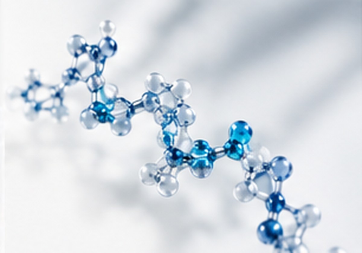
Short peptide fragments of larger hormones are useful for identifying which part of a molecule is responsible for an observed activity. KPV is a classic example: a tripeptide from the C-terminus of α-MSH investigated for signaling effects in cell and animal models.
What Is KPV?
KPV is the tripeptide L-Lys-L-Pro-L-Val (≈342 Da), matching residues 11–13 of α-MSH, a 13-amino-acid peptide derived from pro-opiomelanocortin (POMC). Research materials are synthetic, sometimes with C-terminal amidation matching the parent hormone.
Why Is It Studied?
α-MSH acts through melanocortin receptors and has been studied for roles in pigmentation and immune signaling. KPV is studied to understand whether a minimal fragment can influence inflammatory signaling pathways, and whether such effects depend on melanocortin receptors.
Research Background
Reviews of α-MSH and related tripeptides summarize anti-inflammatory signaling effects observed in vitro and in vivo. A 2008 study reported that KPV is transported into intestinal epithelial cells by the di/tripeptide transporter PepT1 and reduced inflammatory markers in cell and mouse colitis models.
Evidence classification: KPV findings are in vitro and animal. Controlled human clinical data are not established.
Mechanism and Scientific Principles
- Cellular uptake via PepT1 in intestinal epithelial models (in vitro).
- Reduced NF-κB and MAPK pathway activation and pro-inflammatory cytokine expression in cell models (in vitro).
- Some activity reported to be independent of melanocortin receptor binding (in vitro).
Analytical and Experimental Considerations
- Very small peptides elute early in reverse-phase HPLC; method conditions must be adapted to retain them.
- Identity by mass spectrometry, including amidated versus free-acid forms.
- Counter-ions can be a large fraction of mass — see Peptide Purity vs Quantity.
For another small peptide studied in cell biology, see GHK-Cu: Laboratory Research Overview.
Limitations of Current Research
The evidence base is small and predominantly preclinical. Receptor-dependent and receptor-independent mechanisms are not fully resolved.
Frequently Asked Questions
Where does KPV come from?
It corresponds to the C-terminal residues 11–13 of α-MSH, a POMC-derived hormone.
What is PepT1?
A proton-coupled di- and tripeptide transporter expressed in intestinal epithelium; research reports it transports KPV into cells.
Why is KPV hard to analyze by HPLC?
Its small, polar structure causes weak retention on standard reverse-phase columns, requiring adjusted methods.
Scientific References
- Dalmasso G, Charrier-Hisamuddin L, Nguyen HT, et al. PepT1-mediated tripeptide KPV uptake reduces intestinal inflammation. Gastroenterology. 2008;134(1):166-178. [In vitro / animal] View source (opens in new tab)
- Brzoska T, Luger TA, Maaser C, Abels C, Böhm M. α-Melanocyte-stimulating hormone and related tripeptides: biochemistry, antiinflammatory and protective effects in vitro and in vivo, and future perspectives for the treatment of immune-mediated inflammatory diseases. Endocr Rev. 2008;29(5):581-602. [Review] View source (opens in new tab)
FuelX Research Material
Related Research

GHK-Cu: Laboratory Research Overview
The copper-binding tripeptide GHK: its chemistry, the cell-culture and gene-expression research behind it, and the gap between laboratory and human evidence.
Read Research Guide: GHK-Cu: Laboratory Research Overview
HPLC Testing Explained
How reverse-phase HPLC separates peptides, how to read a chromatogram, and what makes a purity method reliable.
Learn More: HPLC Testing Explained
Peptide Purity vs Quantity
Why a 99%-pure peptide can still contain less peptide than the gross weight suggests — and how net peptide content is determined.
Learn More: Peptide Purity vs QuantityContinue Your Research
Read our testing standards to see how each compound is verified.